Themes

Using nanodiamonds to understand the inner workings of the body
Light-based imaging and sensing tools can assist with our understanding of the complex chemical and molecular processes taking place in and around cells in the living body. Fluorescent nanodiamonds (NDs) are an attractive nanoscale-tool that have a range of unique properties which make them highly desirable for bioimaging and biosensing applications. Their fluorescence is produced via optical excitation of atomic defects, such as the negatively charged nitrogen vacancy centre, within the diamond crystal lattice. Possessing long-wavelength emission, high brightness, no photobleaching, no photoblinking, nanometer size, a room temperature sensitivity to magnetic and microwave fields, and an exceptional resistance to chemical degradation make NDs almost the ideal fluorescent bioimaging nanoprobe.
Contact: Brant Gibson and Andrew Greentree

Laser Threshold Magnetometry
Ultra-precise sensing of magnetic fields is of important for the sensing of neuronal activity in the brain (magneto-encephalography), geomagentic sensing, and defence applications. We have discovered a new way to sense magnetic fields using ensembles of nitrogen-vacancy (NV) colour centres in diamond. This approach uses the NV centres as a laser medium, and promises femto-Tesla per root Herz sensitivity at room temperature.
Contact: Andrew Greentree

Quantum enhanced imaging
Most approaches to microscopy implicitly only use the classical (wavelike) nature of light. However, the quantum (particle-like) nature of light also provides information that classical approaches miss. Our theoretical treatment has shown new approaches show how photon counting can boost the resolution of conventional microscopes without increasing the overall photon budget.
Contact: Andrew Greentree
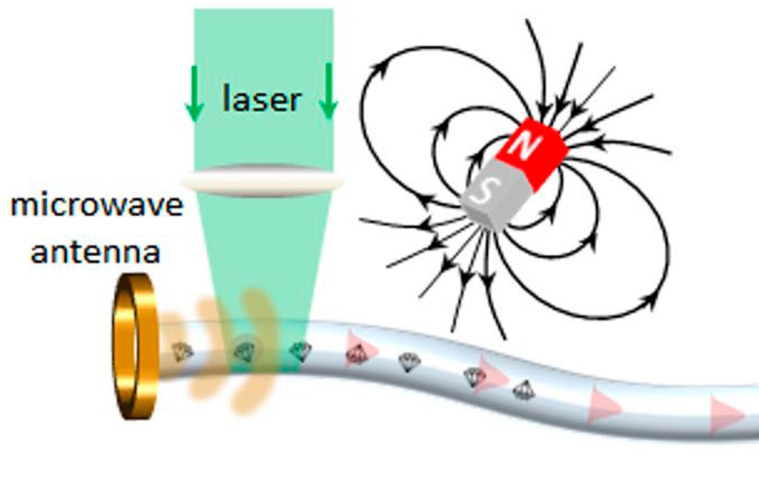
Diamond in fibre sensing
Over the past decade, we have developed techniques to embed fluorescent nanodiamond particles directly into optical fibers. Using novel integrations techniques, such as glass volume-doping and interface doping, diamond particles can be embedded within the glass at the melting and fiber drawing stage, respectively. In collaboration with colleagues, we are exploring this approach to create a fibre-optic sensing platform which is intrinsically sensitive to magnetic fields with remote optical readout.
Contact: Brant Gibson and Andrew Greentree

Diamond hybrid materials
Sometimes, diamond can be used to enhance the properties of other, more conventional materials. In collaboration with our partners, we have been exploring diamond+titanium for biomedical implant technology, diamond+silk for sensing and drug delivery, and diamond + PCL for smart tissue scaffolding. In most cases, diamond preserves, or even enhances the biocompatibility of the original material, and introduces novel imaging and sensing modalities. Lastly, we are working to ensure that the resultant hybrid materials are compatible with existing fabrication technologies such as 3D printing and electrospinning.
Contact: Brant Gibson and Andrew Greentree

Fibre bundle 3D endoscopy
Optical fibre bundle microendoscopes are widely used for visualizing hard-to-reach areas of the human body. These ultrathin devices often forgo tunable focusing optics because of size constraints and are therefore limited to 2D imaging modalities. Ideally, microendoscopes would record 3D information for accurate clinical and biological interpretation, without bulky optomechanical parts. In our research, we have demonstrated that the optical fibre bundles commonly used in microendoscopy are inherently sensitive to depth information. We use the mode structure within fiber bundle cores to extract the spatio-angular description of captured light rays—the light field—enabling digital refocusing, stereo visualization, and surface and depth mapping of microscopic scenes at the distal fibre tip. Our approach is resilient to fibre bending, making it attractive for a wide range of applications.
Contact: Brant Gibson

Theory
We perform a range of theory and modelling tasks relating to electromagnetic and plasmonic modelling, photoacoustic coupling, waveguiding, and quantum simulation.
Contact: Andrew Greentree

Multimodal imaging
Multimodal imaging, a combination of imaging modalities, can provide richer information and overcome the limitations of the independent imaging techniques. This approach has the potential to provide profound new information from a broad range of samples ranging from biological to solid-state materials and photonic devices. Imaging modalities such as Coherent anti-Stokes Raman spectroscopy (CARS), Stimulated Raman spectroscopy (SRS) and Fluorescence-lifetime imaging microscopy (FLIM) are being combined with two photon, confocal fluorescence, and quantum microscopy. This broad range of multimodal imaging techniques can examine label-free vibrational signatures in materials, characterise individual atoms with optical signatures, and be used to unravel the spectral fingerprint of molecules. This powerful imaging tool kit will unlock new knowledge across the fields of physics, chemistry, biology, and beyond.
Contact: Brant Gibson